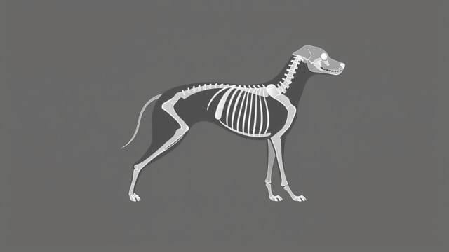
When a beloved dog named Malachi needed medical evaluation, the vet recommended an X-ray to assess internal health. An X-ray, also known as radiography, uses safe, low-level radiation to capture images of the inside of the body. In Malachi’s case, it provided a clear view of bones, joints, lungs, and abdomen. This non-invasive procedure can be performed quickly, usually with the dog gently sedated or calm, allowing veterinarians to diagnose fractures, arthritis, tumors, foreign objects, or respiratory issues. The process is painless and can be completed in minutes, helping guide treatment decisions for dogs like Malachi and ensuring prompt and accurate care.
Why X-rays Are Important for Dogs Like Malachi
X-rays are critical diagnostic tools in veterinary medicine. For Malachi, they offered essential insights into his condition without surgery. They helped detect injuries or illnesses such as:
- Bone fractures or joint abnormalities
- Arthritis in aging pets
- Foreign objects swallowed or lodged
- Lung issues including pneumonia or tumors
- Bladder stones or gastrointestinal blockages
These immediate insights empower veterinarians to make informed decisions about treatment, surgery, or medication plans.
How Malachi’s X-ray Appointment Was Prepared
Prior to imaging, Malachi’s veterinary team prepared carefully to ensure safety and accuracy. Key steps included:
- A health check to ensure Malachi could undergo sedation if required
- Minimal fasting before abdominal X-rays to reduce stomach content
- Gentle sedation or calming techniques to keep Malachi still
- Proper positioning in the X-ray room using supportive pads and foam wedges
These preparations help ensure clear, diagnostic-quality images while prioritizing Malachi’s comfort.
Typical X-ray Views Captured
Veterinarians commonly take several standard views depending on the clinical concern:
1. Lateral View
Taken from the side of the dog, this view reveals the spine, rib cage, lungs, and abdominal organs. It helps assess alignment, fluid in lungs, and masses.
2. Ventrodorsal (VD) or Dorsoventral (DV) View
These top-down or bottom-up views show chest and abdominal organs and are essential for diagnosing issues like heart enlargement or bladder stones.
3. Focused Joint or Limb Views
If Malachi had limping or suspected fractures, targeted shots of specific limbs or joints would be captured such as elbow, hip, or carpus.
Interpreting Malachi’s X-rays
After imaging, a veterinarian or veterinary radiologist reviewed Malachi’s X-rays for signs of injury or disease. They examined:
- Bone structure for fractures, dislocations, or deformities
- Joint space narrowing or bone spurs indicating arthritis
- The presence of gas, fluids, or foreign objects in the abdomen
- Signs of lung disease, such as opacities or masses
These evaluations help determine whether treatments like surgery, physiotherapy, medication, or further diagnostics are needed.
Common Findings in Dog X-rays
Depending on Malachi’s presenting symptoms, his X-rays might have revealed various conditions:
Bone Fractures or Trauma
Broken bones appear as sharp disruptions in bone continuity, allowing vets to plan for surgical repair or splinting.
Arthritis or Degenerative Joint Disease
Older dogs often show narrowed joint spaces and spurs, leading to recommendations for pain relief, supplements, or exercise modification.
Foreign Bodies
Swallowed objects like sticks, stones, or toys can show up clearly and require removal via endoscopy or surgery.
Abnormal Masses or Tumors
Soft tissue masses may appear as shadowy areas requiring biopsy or further imaging like ultrasound.
Respiratory Conditions
X-rays can reveal signs of pneumonia, fluid in lungs, heart enlargement, or other thoracic issues.
Treatment Paths for Findings
Based on what showed up in Malachi’s X-rays, possible next steps included:
- Fracture repair through surgery or splints
- Medications like NSAIDs for arthritis
- Removal of foreign objects surgically or via endoscope
- Antibiotics or supportive care for lung infections
- Further imaging or biopsy for abnormal masses
These targeted treatments help ensure Malachi received precise and effective care.
Advantages and Considerations of X-ray Imaging
X-rays in veterinary care offer many benefits:
- Quick and non-invasive diagnostic tool
- Sensitive to bone conditions and many visible soft tissue abnormalities
- Widely available and reasonably priced
However, it’s important to consider:
- Limited soft tissue contrast compared to ultrasound or CT
- Need for sedation if the dog won’t stay still
- Minor radiation exposure, which vets minimize through safety protocols
Follow-up After Malachi’s X-ray
After his imaging, Malachi’s vet likely discussed findings with his owners and recommended follow-up steps. Options included:
- Prescriptions for pain relief or inflammation
- Rest or restricted activity during healing
- Follow-up X-rays or ultrasound to track progress
- Referral to a specialist for complex conditions
Clear communication ensured his owners understood care recommendations and what to monitor at home.
Preparing Your Dog for an X-ray
If your dog needs an X-ray, here are some tips to make the process smoother:
- Follow any fasting directions for abdominal imaging
- Bring comfort items like a familiar blanket or favorite toy
- Use calming pheromones or mild sedation if advised
- Ask questions about the procedure and anticipated timeline
A well-informed owner and calm pet make for quicker, clearer results.
An X-ray proved invaluable in uncovering Malachi’s internal health status, guiding precise care for any fractures, obstructions, infections, or chronic conditions. This diagnostic tool, combined with the skill of veterinary professionals, ensures dogs like Malachi receive prompt, effective treatment. By demystifying the X-ray process and understanding its potential results, pet owners can be active partners in their dog’s care journey, helping maintain health, comfort, and quality of life over the long term.
: